Measuring Age Itself: The Quiet Revolution in How We Quantify Getting Older
Two new papers — a framework for what a real aging biomarker must do, and a label-free way to read it inside mitochondria — suggest biological age is finally becoming measurable science.
For most of modern medicine, aging has been the variable we could not measure. We could count birthdays, tally diagnoses, and chart the slow drift of cholesterol and creatinine, but the underlying process — the cellular weather that turns a resilient forty-year-old into a frail eighty-year-old — has remained stubbornly invisible. Two recent papers, read side by side, suggest that this is beginning to change. One, a sweeping review in Physiological Reviews, lays out what a credible biomarker of aging must actually do before it earns the name. The other, in Communications Biology, demonstrates a non-destructive way to watch aging unfold inside mitochondria, in living tissue, at subcellular resolution. Together they sketch the outline of a discipline that is starting to grow up.
- The bar is being raised. A new framework argues that aging biomarkers must predict morbidity and mortality, not just correlate with chronological age.
- Function still leads molecules. Today, measures of resilience and frailty have the strongest human evidence; molecular clocks are promising but earlier-stage.
- Mitochondria come into focus. A label-free imaging method reads age-related shifts in NAD(P)H fluorescence inside living cells.
- Evidence is moderate, not settled. The mitochondrial work is in C. elegans; human translation is the open question.
- Implication for readers: Be skeptical of any consumer 'biological age' score that cannot explain what it predicts and in whom.
What counts as a biomarker of aging
The Physiological Reviews piece by Furrer and Handschin is, in effect, a referee's rulebook for a field that has been playing without one. The authors note that aging research has enjoyed an exponential surge in funding and attention, yet much of the resulting evidence remains trapped in model organisms, with human validation hampered by the sheer time it takes for people to age. The bottleneck, they argue, is the absence of biomarkers robust enough to compress decades of biology into measurable windows of months or years — and they set out the criteria such markers would need to meet before clinicians should trust them. You can read the full case in their review.
Their taxonomy is useful because it refuses to flatten the problem. On one side sit molecular candidates — epigenetic clocks, proteomic signatures, transcriptomic patterns — which hold genuine promise but whose predictive value in humans is still being established. On the other side sit the unglamorous incumbents: measures of function, resilience, and frailty, which already have proven predictive value for morbidity and mortality. Grip strength, gait speed, and the ability to recover from a stressor are not as photogenic as a methylation array, but they remain the benchmarks any molecular contender must beat.
A biomarker of aging is not a number that goes up with birthdays. It is a number that tells you something birthdays do not.
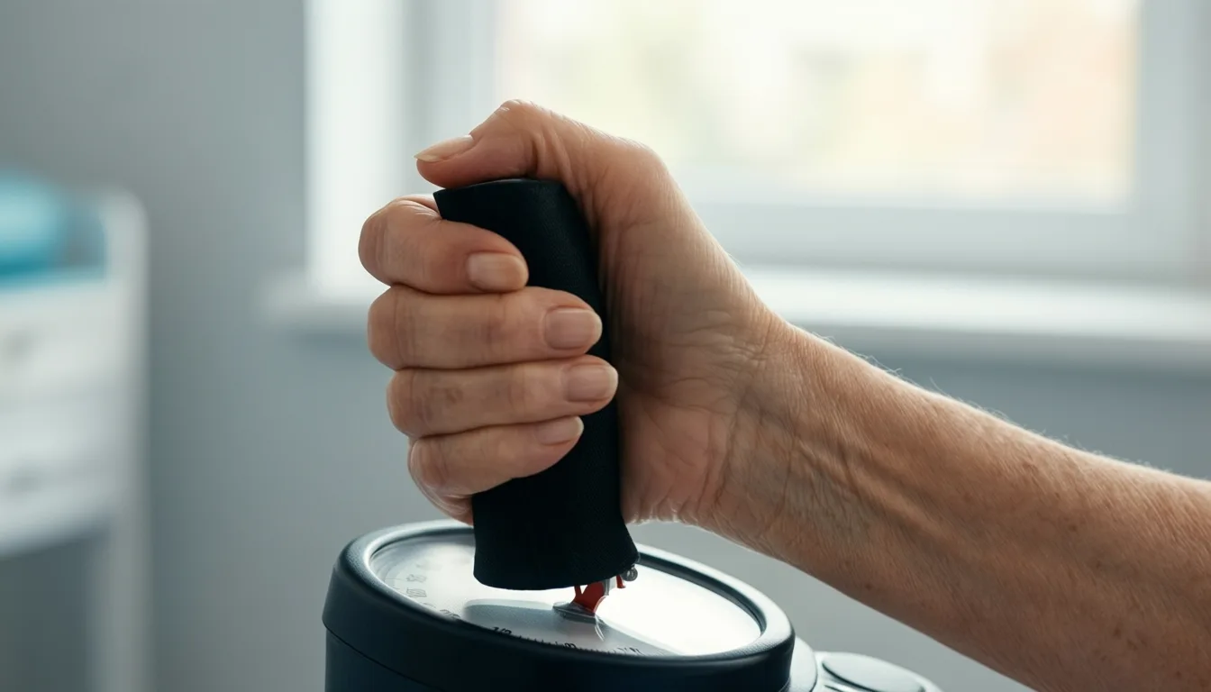
Functional measures like grip strength remain the most validated indicators of aging in humans — the bar that molecular clocks must clear.
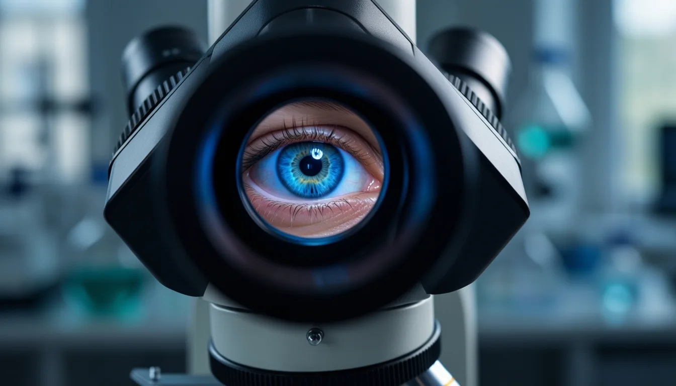
Reading age inside the mitochondrion
If Furrer and Handschin define the goalposts, Morrow and colleagues offer an unusually elegant attempt to reach them. Their study in Communications Biology uses fluorescence lifetime imaging — FLIM — to track the endogenous fluorescence of NAD(P)H inside mitochondria. NAD(P)H is a metabolic cofactor whose biophysical behavior shifts with cellular state; by reading those shifts optically, the team avoids the usual costs of aging assays. The method is non-destructive, label-free, and resolved at the subcellular scale, sidestepping the slow processing times and tissue destruction that limit most existing clocks.
Working in Caenorhabditis elegans, the authors show that mitochondrial NAD(P)H signatures change predictably with age across tissues, track the decline of physiological function, and can be assembled into what they call 'mito-NAD(P)H age clocks'. Crucially, these clocks do more than estimate how old an animal is. They resolve heterogeneity between individuals aging at different rates and, in the paper's most striking claim, predict remaining lifespan. Long-lived worms show a ubiquitous attenuation of the age-related mitochondrial changes — a quieting of the signal, tissue by tissue.

In worms, mitochondrial NAD(P)H fluorescence shifts with age across tissues — and quiets in long-lived individuals.
Why the pairing matters
Read in isolation, each paper is interesting. Read together, they form something closer to a thesis. The review supplies the epistemology — the insistence that an aging biomarker earn its keep by predicting outcomes humans actually care about, not by curve-fitting to the calendar. The imaging study supplies a methodological proof of concept that meets several of those criteria at once: it is quantitative, longitudinal, mechanistically grounded in mitochondrial biology already implicated in aging, and — at least in worms — predictive of the outcome that matters most.
The honest caveats are large. The FLIM clock has been demonstrated in a nematode roughly a millimeter long, not in a human liver or brain. Translating subcellular optical readouts into clinically usable measurements in opaque mammalian tissue is a genuinely hard problem, and the leap from predicting worm lifespan to forecasting human healthspan is the leap the entire field is trying to make. The Physiological Reviews authors are blunt about this gap: model-organism findings have, repeatedly, failed to translate.
What it means for the longevity-minded reader
For readers tracking the cutting edge, the practical takeaway is restraint. The science is moving in the right direction, but the evidence rating here is moderate, not high. The most validated measurements you can act on today remain the ones a good geriatrician has used for decades: how fast you walk, how hard you grip, how well you recover from physiological stress. The molecular and optical clocks now in development may eventually surpass these — and the Morrow paper is a credible early signal — but 'eventually' is doing a lot of work in that sentence.
The smarter posture is to treat the next few years of aging-biomarker news the way Furrer and Handschin implicitly recommend: ask what the marker predicts, in which population, against which functional benchmark, and with what evidence in humans. Anything that cannot answer those four questions is, for now, a hypothesis with good lighting. None of this is medical advice; decisions about testing or treatment belong with a clinician who knows your history.
What is genuinely new is the direction of travel. For the first time, the field has both a rulebook for what a real aging biomarker must do and a working example of a measurement subtle enough to try. That is not a cure, and it is not a clock you can buy. It is something more interesting: the early outline of a medicine that can finally see what it is treating.

Label-free imaging is shifting aging research from destructive endpoint assays to longitudinal observation of living tissue.
Frequently asked questions
What makes something a true biomarker of aging, according to the article?
The article, drawing on the Furrer and Handschin review, argues that a biomarker of aging must predict morbidity and mortality, not simply correlate with chronological age. A number that merely goes up with birthdays does not qualify; it must tell you something birthdays do not.
Why do grip strength and gait speed still matter more than molecular clocks?
The article describes functional measures like grip strength, gait speed, and the ability to recover from a stressor as the field's most validated indicators because they already have proven predictive value for morbidity and mortality in humans. Molecular candidates such as epigenetic clocks are described as promising but earlier-stage, and functional measures set the benchmark any molecular contender must beat.
What is the FLIM method and what did the researchers find with it?
Fluorescence lifetime imaging (FLIM) reads the endogenous fluorescence of NAD(P)H inside mitochondria without dyes, stains, or tissue destruction, at subcellular resolution. Working in C. elegans, the researchers showed that these mitochondrial signatures change predictably with age across tissues and can predict remaining lifespan, with long-lived worms showing an attenuation of the age-related signal.
Why hasn't the mitochondrial clock been proven useful for people yet?
The study was conducted in C. elegans, a nematode roughly a millimeter long, and the article calls translating subcellular optical readouts into clinically usable measurements in opaque mammalian tissue a genuinely hard problem. The article also notes that model-organism findings have repeatedly failed to translate to humans.
What does the article say readers should watch for to know if this science is progressing?
The article identifies three signals: whether any molecular or imaging-based clock prospectively predicts morbidity and mortality in humans as well as grip strength and gait speed already do; whether label-free optical methods can be adapted to mammalian tissues; and whether consumer biological age products begin disclosing what they predict, in whom, and with what confidence intervals.
Sources
- Biomarkers of aging: from molecules and surrogates to physiology and function. — Physiological reviews
- Endogenous mitochondrial NAD(P)H fluorescence can predict lifespan. — Communications biology
Join the conversation
Comments are moderated and reviewed before they appear. Be constructive — this is health information.
Add your comment