
In This Issue


The Disuse Penalty: How Fast Fat Invades Resting Muscle
A new PRISMA-grade systematic review puts numbers on what endurance athletes intuit — even a short layoff lets fat creep into the muscle itself, and that may be the spark for the insulin resistance that follows.
Ask any endurance athlete what they fear about a forced layoff and they will tell you about VO2 max sliding, mitochondria thinning, the cruel first run back. What they rarely mention — because until recently the data were scattered — is the quieter change happening inside the muscle itself. Fat moves in. Not the visible kind around the waistband, but a finer, more consequential kind: droplets wedged between fibers and pooled around them, marbling a tissue that is supposed to be lean. A new systematic review in Experimental Physiology finally puts numbers on how fast that marbling appears when you stop moving, and the numbers are smaller than the tabloid headlines but more interesting than the shrug response.
The review, published in 2026 by Prokopidis, Reyes and Duque, followed PRISMA methodology across PubMed, Scopus, Web of Science and the Cochrane Library, with bias assessed using RoB2 and ROBINS-I. Nine clinical trials in healthy adults met the bar. The question was narrow and useful: when otherwise healthy people undergo bed rest or unilateral lower-limb immobilization, what happens to intramuscular adipose tissue (IntraMAT, the fat stored within the muscle) and intermuscular adipose tissue (InterMAT, the fat sitting between muscle groups beneath the fascia)? Both depots are increasingly viewed as mechanistic players in insulin resistance, not just passive consequences of it.
The headline finding for the postural muscles is the one that should make desk-bound athletes sit up straighter. Bed rest increased IntraMAT in the lumbar multifidus by 18.7%, with smaller but measurable bumps of 5.4% in the erector spinae and 4.5% in the quadratus lumborum. These are the deep stabilizers of the spine — exactly the tissue that endurance athletes rely on to hold form in hour four of a ride or kilometer 35 of a marathon. The lower-limb picture from immobilization studies was more mixed, with InterMAT changes in the thigh that did not always reach statistical significance, but the direction of travel was consistent.
Why marbling matters for metabolism
To appreciate why a few percentage points of fat infiltration is interesting, you have to remember what muscle is supposed to do beyond moving you forward. Skeletal muscle is the body's largest sink for postprandial glucose. Insulin opens the door, GLUT4 transporters surface, and glucose floods in to be burned or stored as glycogen. When lipid metabolites accumulate inside or around fibers, that signaling cascade gets noisier — diacylglycerols and ceramides interfere with insulin receptor substrate phosphorylation, and the door stops opening as cleanly. The authors of the review explicitly frame IntraMAT and InterMAT accumulation as a plausible mechanistic link between disuse and the insulin resistance that reliably appears after bed rest.
The evidence rating here is moderate, and worth holding honestly. Nine trials is a modest pool. Imaging methods varied. Healthy adults are not the same population as a recovering surgical patient or a long-haul traveller, and narrative synthesis cannot deliver the tidy pooled effect size a meta-analysis would. What the review does establish is that the signal is real and reproducible enough to take seriously, and that the postural muscles seem unusually susceptible — perhaps because they are the ones most completely unloaded when you are horizontal.
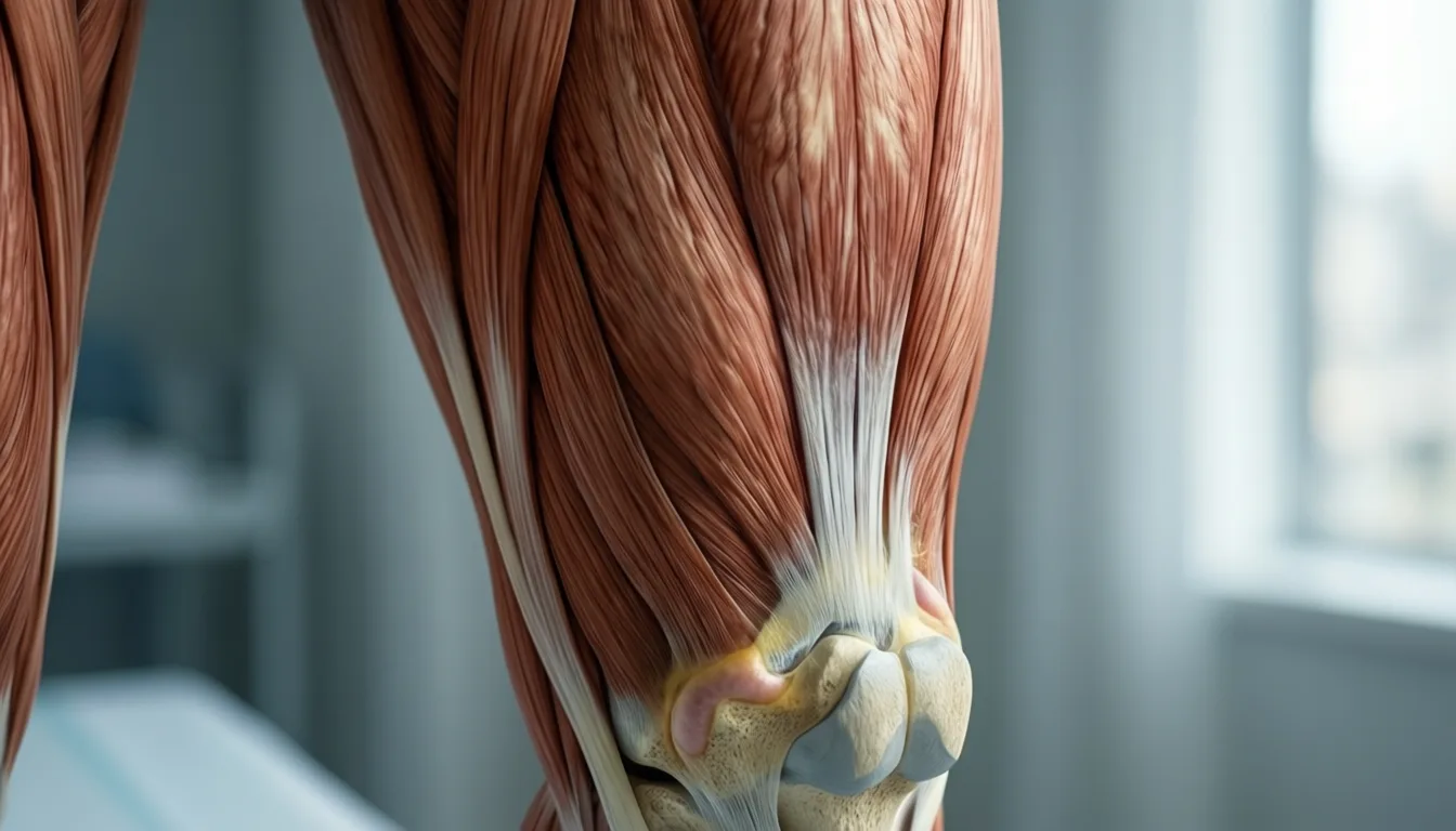
IntraMAT sits within the muscle; InterMAT pools between muscle groups beneath the fascia. Both are visible on MRI and CT, and both rise with disuse.
The deep stabilizers of the spine showed the largest fat infiltration — exactly the tissue endurance athletes rely on to hold form deep into a session.
The mechanistic logic for movement during downtime
The practical reading is not panic. It is permission to take the small stuff seriously. The conditions studied — frank bed rest and limb casting — are extremes, but the directionality is consistent with what's been observed in step-reduction protocols and post-surgical immobilization elsewhere in the literature. If multifidus marbling can shift measurably in days of horizontal stillness, the case for ankle circles on a long-haul flight, a daily walk during a flu, or sub-maximal cycling around a foot injury is not aesthetic. It is mechanistic.
For the performance-minded reader, the more interesting question is whether load-matched movement — even tiny, sub-aerobic doses — can blunt the IntraMAT response while the inflammatory or structural reason for the layoff resolves. The review does not answer this; it sets the table. Future trials with controlled, partial-loading arms are the next move, and the authors point at that gap. For now, the takeaway is that disuse changes muscle composition, not just muscle size, and the composition change is the kind that matters for how your body handles its next meal.
- Disuse changes composition, not just size. Even short bed rest pushes measurable fat into the muscle itself.
- Postural muscles are most exposed. The lumbar multifidus showed an 18.7% IntraMAT rise in pooled bed-rest data.
- The metabolic stakes are real. Intramuscular lipid accumulation is mechanistically linked to insulin resistance.
- Evidence is moderate, not definitive. Nine trials, narrative synthesis, varied imaging — directional but not pooled.
- Movement during downtime is plausible insurance. Even sub-aerobic loading may matter; ask a clinician what's safe for your situation.
The romance of training is in the hard sessions — the threshold intervals, the long runs, the calibrated suffering. But what this review quietly underlines is that the architecture you build in those sessions has a maintenance cost paid in ordinary, unglamorous movement. Muscle is not just contractile machinery; it is a metabolic organ, and its composition is being negotiated, week by week, by how often you ask it to work. The disuse penalty is small per day, and that is exactly why it deserves attention. Small daily things, repeated, are how physiology actually changes.
Frequently asked questions
What is the difference between IntraMAT and InterMAT?
IntraMAT (intramuscular adipose tissue) is fat stored within the muscle itself, while InterMAT (intermuscular adipose tissue) pools between muscle groups beneath the fascia. Both depots are visible on MRI and CT, and both rise with disuse.
Which muscles showed the greatest fat infiltration during bed rest?
The deep postural muscles of the spine were most affected. Bed rest increased IntraMAT in the lumbar multifidus by 18.7%, with smaller but measurable rises of 5.4% in the erector spinae and 4.5% in the quadratus lumborum. Lower-limb immobilization data were more variable and did not always reach statistical significance.
Why does fat accumulating inside muscle matter for metabolism?
Skeletal muscle is the body's largest sink for postprandial glucose, and when lipid metabolites such as diacylglycerols and ceramides build up inside or around fibers, they interfere with insulin receptor substrate phosphorylation, disrupting the signaling that allows glucose to enter the muscle. The review authors explicitly frame IntraMAT and InterMAT accumulation as a plausible mechanistic link between disuse and the insulin resistance that reliably appears after bed rest.
How reliable is the evidence in this review?
The authors rate the evidence as moderate. This is a narrative systematic review of nine clinical trials — not a meta-analysis — imaging methods varied across studies, and findings from healthy adult cohorts may not generalize to older or post-surgical populations where baseline IntraMAT is already higher. The review establishes that the signal is real and reproducible enough to take seriously, but the percentages should be treated as directional rather than definitive.
Does the review show that staying active during a layoff can prevent muscle fat infiltration?
The review does not answer this question directly; it describes that gap as the next area for future trials with controlled, partial-loading arms. The authors do note that the case for sub-maximal movement during downtime — such as ankle circles on a long-haul flight or cycling around a foot injury — is mechanistic, not merely aesthetic, based on the consistent direction of the disuse findings.
Sources

MASLD and Your Kidneys: The Quiet Conversation Between Two Organs
The disease formerly known as fatty liver got a new name in 2023 — and a fresh acknowledgment that it rarely travels alone. Here's what the latest review tells us about the liver-kidney link.
If you have ever tried to read a health headline while holding a toddler on one hip and a lukewarm coffee in the other hand, you already know the rules of engagement: tell me what changed, tell me if it matters, and tell me what to do before someone needs a snack. So here is the short version. In 2023, the medical world quietly renamed the most common chronic liver condition in the world. What used to be called non-alcoholic fatty liver disease is now MASLD — metabolic dysfunction-associated steatotic liver disease. The new name is clunky, but it does something important: it admits, out loud, that this is a whole-body metabolic story, and that one of the organs listening most closely is the kidney.
A new review in the Journal of Clinical and Translational Hepatology pulls together what researchers currently understand about how MASLD and chronic kidney disease (CKD) travel together — epidemiologically, biologically, and clinically. The short read: the two conditions share so much of the same metabolic plumbing that treating one without thinking about the other is starting to look like an oversight. The longer read is more careful, because the evidence here is moderate, not airtight.
For parents juggling pediatrician visits and their own postponed checkups, this matters less as a diagnosis to fear and more as a frame to keep in mind. Metabolic health is a system, not a single number on a lab slip.
Why the name change actually matters
The old label — non-alcoholic fatty liver disease — defined the condition by what it wasn't. The new criteria flip that. MASLD is diagnosed when hepatic steatosis (fat in the liver) shows up alongside cardiometabolic risk factors like elevated blood sugar, blood pressure, waist circumference, or lipid abnormalities. In other words, the diagnosis now points directly at the metabolic context the liver is sitting in.
That framing matters because it lines up with how the disease actually behaves in the body. MASLD and CKD have shown a significant global increase in comorbidity, driven largely by the rise of metabolic syndrome. They are not separate stories happening to the same people by coincidence.
The new name codifies what clinicians have long suspected: fatty liver rarely shows up alone.
What the review found about risk
The headline finding, stated carefully: MASLD is an independent risk factor for chronic kidney disease, and the risk appears to track with the severity of liver fat and the progression of hepatic fibrosis. The more advanced the liver disease, the more the kidney signal shows up alongside it.
The relationship also seems to run in both directions. CKD may itself be a risk factor for fibrosis progression in people with MASLD, which suggests a feedback loop rather than a one-way street. When the two coexist, the review notes, the interaction may accelerate cardiovascular events and increase all-cause mortality risk.
A note on language: "may," "appears to," and "associated with" are doing real work in those sentences. The authors themselves flag that the bidirectional causal relationship — which condition is truly driving the other, and through what molecular conversation — remains unclear. This is a moderate-evidence story, not a settled one.
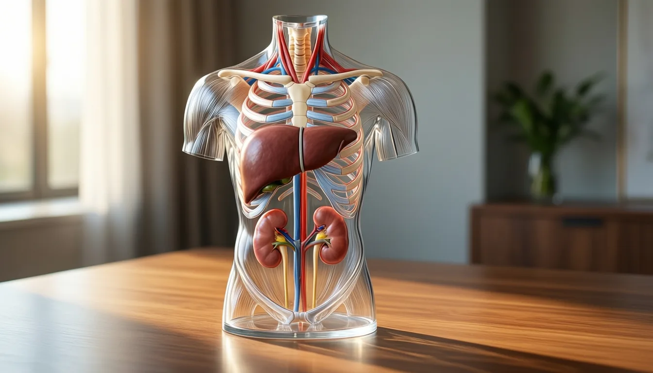
Metabolic health is a system, not a single number on a lab slip.
The shared machinery
Why would a liver problem and a kidney problem keep showing up at the same dinner table? Because they appear to be eating from the same plate. The review identifies several core pathophysiological mechanisms MASLD and CKD share:
- Genetic variants that influence how the body handles fat and sugar.
- Insulin resistance, the metabolic background hum of much modern chronic disease.
- Lipid metabolism disorders — the body's fat-handling system getting out of tune.
- Chronic low-grade inflammation, the kind that doesn't make you feel sick but quietly remodels tissue over years.
- Oxidative stress, an imbalance between cellular damage and repair.
- Gut microbiota dysbiosis — shifts in the bacterial communities that help regulate metabolism and inflammation.
None of these is unique to either organ. That is precisely the point. When the underlying metabolic environment tilts, multiple downstream systems tilt with it.
Everyday meals are still where most of the metabolic conversation happens.
- New name, same disease, sharper focus. MASLD replaces the old NAFLD label and explicitly ties fatty liver to cardiometabolic risk factors.
- Liver and kidneys are linked. MASLD is an independent risk factor for CKD, and severity tracks with liver fibrosis.
- The relationship looks bidirectional. CKD may, in turn, accelerate liver fibrosis — though causality is not yet nailed down.
- They share the same plumbing. Insulin resistance, inflammation, lipid handling, and gut microbiome shifts show up in both.
- The combination raises the stakes. Together, MASLD and CKD appear to push up cardiovascular and mortality risk.
- Evidence is moderate, not final. Clinical prediction tools and targeted treatments for the overlap are still works in progress.
What this means at the kitchen table
If you are running on four hours of sleep and a granola bar, here is the kind, realistic version. You do not need to memorize a new acronym or overhaul your life this week. You do not need to panic if a past blood test mentioned a fatty liver. What this body of research is gently arguing is that the levers that help one organ in this system tend to help the others: steady blood sugar, healthy blood pressure, a waistline that is not creeping, sleep that exists at all.
For a tired parent, the smallest useful step is almost always the right one. A walk after dinner. A glass of water before the second coffee. A real meal instead of finishing your kid's crusts standing up. Booking the physical you have rescheduled three times. None of that is glamorous. All of it is the same medicine the liver and the kidneys are quietly asking for.
Where the science still has homework
The review is candid about its own limits. There are significant gaps in clinical prediction tools and in targeted treatment strategies for people who have both conditions. The molecular dialogue between the organs is still being mapped. For readers, that means two things at once: this is a real and increasingly recognized clinical pattern, and the precise playbook for managing it is still being written.
That is an honest place to land. Not a miracle, not a panic — a clearer picture of a metabolic system that has always been talking to itself, and a name that finally reflects it.
Frequently asked questions
What does MASLD stand for, and why did the name change from non-alcoholic fatty liver disease?
MASLD stands for metabolic dysfunction-associated steatotic liver disease, a label adopted in 2023 to replace the old non-alcoholic fatty liver disease (NAFLD). The new name points directly at the metabolic context surrounding the diagnosis — hepatic steatosis plus cardiometabolic risk factors such as elevated blood sugar, blood pressure, waist circumference, or lipid abnormalities — rather than defining the condition by what it isn't.
Is MASLD a risk factor for chronic kidney disease, or is the link just a coincidence?
According to the review covered in the article, MASLD is an independent risk factor for chronic kidney disease, and the risk appears to track with the severity of liver fat and the progression of hepatic fibrosis. The two conditions have also shown a significant global increase in comorbidity, driven largely by the rise of metabolic syndrome, suggesting the connection is not coincidental.
Does chronic kidney disease affect the liver in return, or does the relationship only run one way?
The relationship appears to run in both directions. The review notes that CKD may itself be a risk factor for fibrosis progression in people with MASLD, pointing to a feedback loop rather than a one-way street. However, the article is careful to note that which condition is truly driving the other remains unclear.
What biological mechanisms do MASLD and chronic kidney disease share?
The review identifies several overlapping mechanisms, including insulin resistance, lipid metabolism disorders, chronic low-grade inflammation, oxidative stress, gut microbiota dysbiosis, and genetic variants that influence how the body handles fat and sugar. Because none of these is unique to either organ, a tilt in the underlying metabolic environment can affect both simultaneously.
How solid is the evidence connecting MASLD and kidney disease — is this settled science?
The article describes the evidence as moderate, not airtight. The authors of the review themselves flag that the bidirectional causal relationship between the two conditions remains unclear, and the article notes that clinical prediction tools and targeted treatment strategies for people who have both conditions are still works in progress.
Sources
- Metabolic Dysfunction-associated Steatotic Liver Disease and Chronic Kidney Disease: From Epidemiology and Pathophysiology to Clinical Prediction and Treatment Options. — Journal of clinical and translational hepatology

Sarcopenia Might Be an Inflammation Story — and That Changes Everything
Two new papers reframe age-related muscle loss as a downstream effect of chronic inflammation, immune aging, and a restless gut. Protein shakes alone won't cut it.
Okay, real talk: I used to think muscle loss in older age was basically a protein problem. Eat more chicken, lift heavier, the end. Then I started reading the new sarcopenia research and had to rebuild my whole mental model. Two 2026 papers — one a sweeping pharmacology review, one a hospital cohort study — keep pointing at the same surprising culprit. It's not just the protein on your plate. It's the inflammation in your background noise.
Quick gloss before we go further: sarcopenia is the age-related loss of muscle mass, strength, and function. It's the reason a grandparent can't open a jar, or why a fall in your 70s turns into a long hospital stay. For years, the standard playbook has been protein plus resistance training. Both still matter — a lot. But researchers are increasingly arguing that those interventions work partly because they push back against something deeper going on in the body.
That "something deeper" has a nickname in the literature: inflammaging. It's the slow, low-grade inflammation that quietly turns up as we get older — not the sharp, useful kind that fights a cold, but a persistent background hum. A 2026 review in Frontiers in Pharmacology lays out the case that this hum is a driving factor behind muscle loss, not a side effect of it.
The five-way pileup inside an aging muscle
Here's the part I had to read twice. The review describes chronic inflammation as the crossroads where several aging processes collide: cellular senescence (cells that refuse to die and keep spitting out inflammatory signals), immunosenescence (an immune system that's both tired and twitchy), oxidative stress, mitochondrial dysfunction (your muscle cells' little power plants sputtering), and gut microbiota dysbiosis — an out-of-whack gut ecosystem leaking inflammatory signals into the bloodstream. Picture five roads merging into one intersection, and the intersection is your quadriceps. That's the model the authors propose.
Obesity complicates it further. Because fat tissue is itself inflammatory, carrying excess weight while losing muscle creates sarcopenic obesity — a combo that the review notes makes both the muscle loss and the functional decline worse.

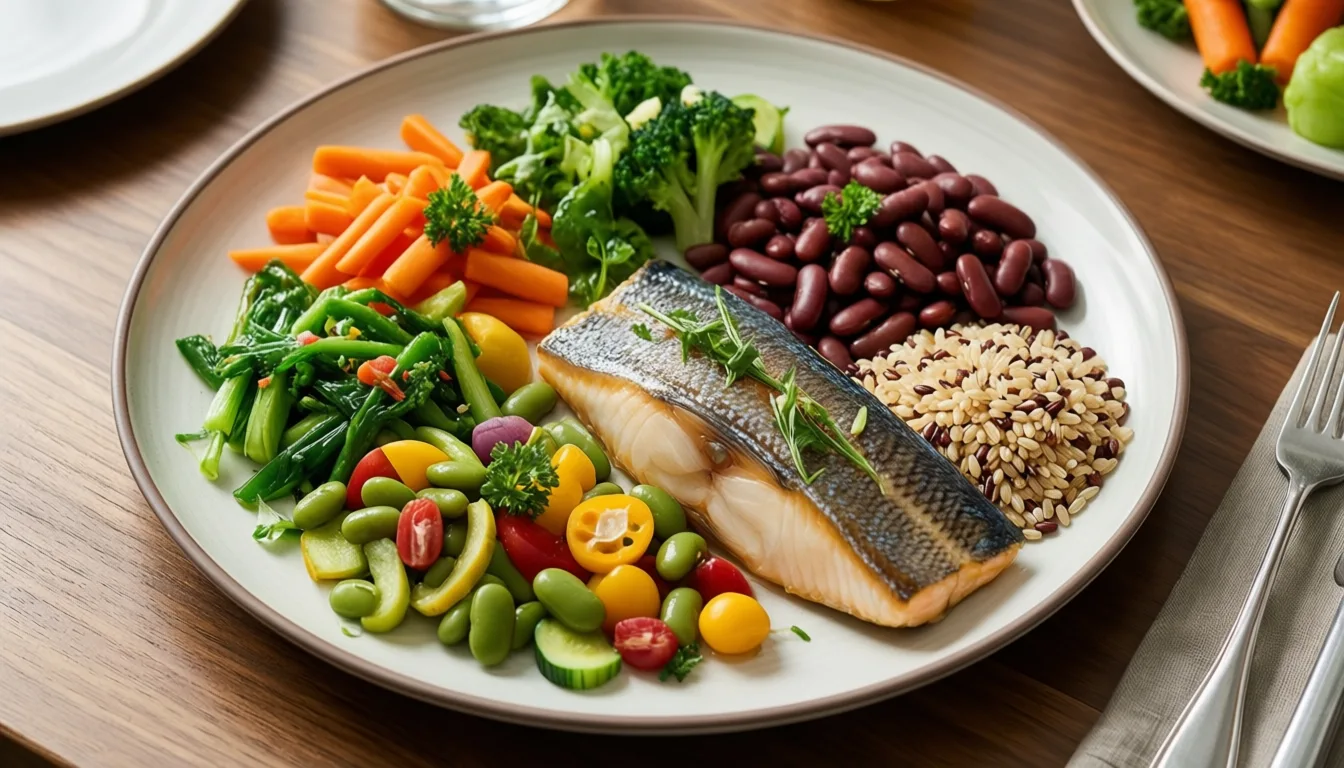
Anti-inflammatory eating patterns are part of the emerging sarcopenia conversation — though the review stops short of endorsing any single diet.
The intervention target is shifting from protein on the plate to the inflammatory weather in the body.
The hospital data that made me sit up
The mechanism story is interesting on its own, but a second 2026 study gives it a real-world edge. Researchers behind the BACK-UPUG cohort looked at 590 older patients moving through a Post-Emergency Geriatric Unit at a French university hospital. The question was simple: who ends up stuck in the hospital longer? Median age was 88. About 42% had a prolonged stay of six days or more.
Two things jumped out. A higher burden of chronic illness raised the odds of a prolonged stay (Charlson Comorbidity Index odds ratio 1.10). And elevated CRP above 64 mg/L — C-reactive protein, a standard blood marker of inflammation — was associated with roughly twice the odds of getting stuck (OR 2.07). Frailty markers tracked alongside it. In other words, the body's inflammatory tone wasn't just a lab curiosity. It predicted who went home and who didn't.
So what actually moves the needle?
Here's where I want to be careful, because the evidence rating for this whole reframe is moderate, not slam-dunk. The pharmacology review is a synthesis of mechanisms and emerging targets, not a prescription. The hospital study is retrospective and observational — it shows association, not that lowering CRP causes shorter stays.
That said, the review is clear about what already has evidence behind it: exercise (especially resistance training), nutritional supplementation, and combined approaches improve muscle mass and function — and bring measurable anti-inflammatory benefits as a bonus. Beyond the classics, the authors flag anti-inflammatory drugs and treatments aimed at inflammatory pathways as a promising frontier. Promising, not proven. That distinction matters.

Resistance training remains the most evidence-backed lever — and it appears to do double duty against inflammation.
- The frame is shifting. Sarcopenia is being described less as a protein-deficit problem and more as a downstream effect of chronic, low-grade inflammation.
- Five pathways converge. Cellular senescence, immunosenescence, oxidative stress, mitochondrial dysfunction, and gut dysbiosis all feed inflammaging.
- Inflammation shows up in outcomes. In a 2026 hospital cohort, CRP above 64 mg/L doubled the odds of a prolonged geriatric stay.
- Exercise still wins. Resistance training and nutrition remain the best-supported interventions — and they have anti-inflammatory effects of their own.
- Drug strategies are early. Anti-inflammatory pharmacology for sarcopenia is a promising lane, not a proven one.
- Talk to a clinician. CRP, frailty scores, and muscle assessments aren't DIY tools — bring questions to a doctor who knows your history.
The takeaway for the rest of us
If you're decades away from a geriatric ward, none of this is meant to scare you. The honest read is that the levers most of us already know about — moving heavy things, eating enough protein, sleeping, not smoking, keeping a reasonable weight — are looking even better because they appear to act on inflammation too. The newer ideas about senescent cells, microbiome tweaks, and targeted anti-inflammatories are exciting, but they're still being figured out.
So no, you don't need to bio-hack your cytokines this weekend. But the next time someone tells you muscle loss is just about eating more chicken, you'll know the story is bigger — and a lot more interesting — than that.
Frequently asked questions
What is inflammaging, and why does it matter for muscle loss?
Inflammaging is the slow, low-grade inflammation that quietly increases as we get older — not the sharp, useful kind that fights infection, but a persistent background hum. A 2026 review in Frontiers in Pharmacology argues this chronic inflammation is a driving factor behind sarcopenia, not merely a side effect of it.
What five biological pathways does the research link to inflammaging and sarcopenia?
The review identifies cellular senescence, immunosenescence, oxidative stress, mitochondrial dysfunction, and gut microbiota dysbiosis as the five converging pathways. Each feeds into the chronic inflammatory environment that the authors propose underlies age-related muscle loss.
What did the French hospital study find about inflammation and length of hospital stay?
In a cohort of 590 older patients with a median age of 88, elevated C-reactive protein above 64 mg/L was associated with roughly twice the odds of a prolonged hospital stay of six or more days. About 42% of patients in the study experienced such a prolonged stay.
Does the new inflammation research mean resistance training and protein are no longer important for sarcopenia?
No — the article is explicit that resistance training and nutritional supplementation remain the best-supported interventions for improving muscle mass and function. The emerging finding is that these approaches also carry measurable anti-inflammatory benefits, suggesting they may work partly by addressing the underlying inflammatory environment.
How strong is the evidence for anti-inflammatory drugs as a sarcopenia treatment?
The article describes anti-inflammatory pharmacology for sarcopenia as a promising frontier, not a proven one, and draws a clear distinction between the two. The pharmacology review it cites is a synthesis of mechanisms and emerging targets rather than a clinical prescription.
Sources
- Chronic inflammation as a driving factor for sarcopenia: an update on pathophysiology and future therapeutic targets. — Frontiers in pharmacology
- Frailty and inflammation predict prolonged stay in post-emergency geriatric units: a retrospective cohort study. — The journal of nutrition, health & aging