Your Retinas May Be Aging Faster Than You Are — and Multimorbidity Follows
A large deep-learning study suggests the gap between your retina's biological age and your calendar age tracks with chronic disease risk. The eye, it turns out, may be a useful longevity dashboard.
Your calendar age is a stubborn number. Your biology is more negotiable — and increasingly, more measurable. Among the newest candidates for a practical longevity readout is the retina, the thin sheet of neural tissue at the back of the eye that doubles as a window onto the body's small vessels and nerves. A deep-learning analysis of 45,436 middle-aged and older adults, published in GeroScience, reports that the gap between a person's retina-predicted age and their actual age is associated with both current chronic-disease burden and the future onset of multimorbidity. The finding is not a verdict on any one reader's health. It is, however, a useful reframing: the eye may be one of the more honest mirrors we have.
- The metric: 'Retinal age gap' is how much older (or younger) a deep-learning model judges your retina to be versus your calendar age.
- The signal: In a study of 45,436 adults, people with one or two-plus age-related conditions had measurably larger retinal age gaps at baseline than those with none.
- The forecast: Each 5-year increase in retinal age gap was linked to an 8% higher risk of developing multimorbidity over a median 11.4 years of follow-up.
- The caveat: This is an association in one large cohort, not a clinical test you can act on yet. Evidence strength is moderate.
- The use case: Treat retinal imaging as one possible future input to whole-person risk — not a diagnosis, and not a reason to chase a number.
Why the eye keeps showing up in longevity research
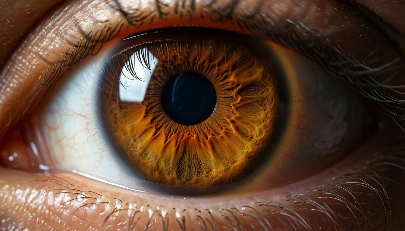
The retina is the only place in the body where clinicians can directly observe small arteries, veins and nerve fibers without cutting anything open. Those microvessels respond to the same pressures, glucose excursions and inflammatory traffic that age the rest of the cardiovascular and neurological system. That is why retinal photographs have quietly become a favored training set for biological-age models: the images are standardized, abundant, and tied to long follow-up data in biobanks.
The GeroScience team applied a previously developed deep-learning model that estimates 'retinal age' from a fundus photograph, then subtracted each participant's calendar age to produce a retinal age gap. They then asked a deceptively simple question: does that gap track with how many chronic diseases a person has, and with how many they go on to develop? In the fully adjusted analysis, the answer was yes on both counts.
A standard fundus image. The same photographs that screen for diabetic eye disease are now training grounds for biological-age models.
What the numbers actually say
At baseline, participants who already carried one age-related condition had a retinal age gap roughly 0.20 years larger than peers with none; those with multimorbidity — two or more conditions — had a gap about 0.25 years larger. Both differences were statistically significant in the fully adjusted model, with confidence intervals that excluded zero. These are population-level averages, not personal verdicts. But they are consistent with the idea that disease burden and retinal aging move together.
The prospective piece is the more interesting one. Over a median follow-up of 11.38 years, 3,607 participants — 17.3% of the at-risk group — developed multimorbidity. Each five-year increment in baseline retinal age gap was independently associated with an 8% higher hazard of incident multimorbidity (HR 1.08, 95% CI 1.02–1.14). 'Independently' is doing real work in that sentence: the association held after adjustment for the usual suspects, which is what elevates this from curiosity to signal.
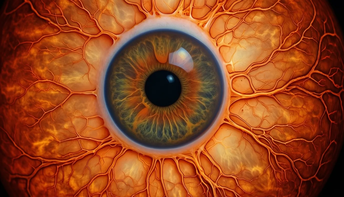
The retina is the only place in the body where a clinician can directly observe small vessels and nerves without an incision. It was always going to end up on the longevity dashboard.
What this is not
This is one large observational cohort with a single retinal-age model and a single composite outcome. It does not show that intervening on the retina — or on anything that nudges the gap — changes disease trajectories. It does not establish that retinal age gap outperforms simpler clinical inputs like blood pressure, HbA1c or a lipid panel. And it cannot tell an individual reader whether their own retina is aging quickly; that would require the model, the image and a clinical context none of us can self-administer.
The evidence rating here is moderate for good reasons. The sample is large and the follow-up long, which is rare and valuable. But the model is one of several in the field, and external validation across populations, cameras and ethnicities remains an open question. Effect sizes are meaningful at population scale and modest at the individual level — an 8% hazard increase per five-year gap is a signal, not a sentence.
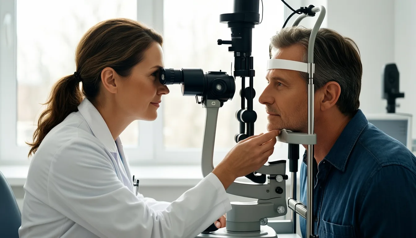
Fundus imaging takes seconds and no dilation in many modern devices. The bottleneck is not the camera — it is the interpretation layer behind it.
How to think about it on a packed calendar
For the executive reader, the practical takeaway is restraint. You do not need a retinal age gap reading to act on what this study reinforces: the same metabolic and vascular forces that age the rest of you also age your retina, and they are largely the levers you already know — sleep, movement, blood pressure, glucose, lipids, stress load. Retinal imaging may eventually sharpen those conversations with a clinician. It does not replace them.
If you already see an optometrist or ophthalmologist annually, ask whether they retain your fundus images and whether their device captures them in a format compatible with future analysis. That is a quiet, no-cost form of optionality. If you are being worked up for cardiometabolic risk, mention that retinal microvascular signals are an active area of research; it may inform how your clinician interprets a borderline finding. Beyond that, the honest advice is to wait for validation and not to chase a number that is not yet yours to chase.
The deeper story the GeroScience analysis tells is not about eyes. It is about the slow normalization of biological age as a clinical concept — a shift from treating chronological age as destiny to treating it as one variable among many. The retina is a convenient proxy because it is photographable, standardized and richly vascular. Other organs will get their own clocks. The reader's job, for now, is to recognize the direction of travel without overreacting to any single instrument on the dashboard.
Frequently asked questions
What exactly is a 'retinal age gap'?
A retinal age gap is the difference between the age a deep-learning model estimates your retina to be — based on a fundus photograph — and your actual calendar age. A positive gap means your retina appears older than you are; a negative gap means it appears younger.
What did the study find about retinal age gap and future disease risk?
Over a median follow-up of 11.38 years, each five-year increase in baseline retinal age gap was associated with an 8% higher hazard of developing multimorbidity. In that period, 17.3% of the at-risk group went on to develop two or more age-related chronic conditions.
Why do researchers use the retina to study biological aging?
The retina is the only place in the body where small arteries, veins, and nerve fibers can be directly observed without any incision. Those microvessels respond to the same pressures, glucose changes, and inflammation that age the cardiovascular and neurological systems, making retinal photographs a useful training set for biological-age models.
Is this a test I can take now to find out how fast my retina is aging?
No — the article describes this as an association from one large observational cohort, not a clinical test you can act on yet. External validation across different cameras, populations, and ethnicities is still an open question, and the evidence strength is rated as moderate.
What practical step, if any, does the article suggest regarding retinal imaging?
The article suggests asking your optometrist or ophthalmologist whether they retain your fundus images and whether their device captures them in a format compatible with future analysis — described as a quiet, no-cost form of optionality. Beyond that, it advises waiting for further validation rather than trying to chase a retinal age gap number.
Join the conversation
Comments are moderated and reviewed before they appear. Be constructive — this is health information.
Add your comment